What is Stroke Volume?
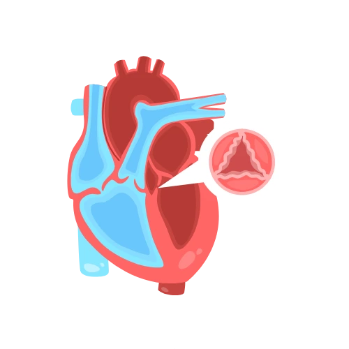
Stroke volume is the volume of blood ejected by the left ventricle with each heartbeat. It is a core determinant of cardiac output and tissue perfusion, and it is routinely measured or estimated in echocardiography, critical care, and cardiology. This calculator supports four methods: LVOT Doppler (the most accurate non-invasive approach), Teichholz M-mode, cardiac output divided by heart rate, and end-diastolic volume multiplied by ejection fraction. By selecting the method that matches your available data, you receive a consistent stroke volume value, an indexed value adjusted for a reference body surface area, and a category that ranges from low to high. The tool is intended for clinicians, sonographers, and students who need a quick, reproducible estimate. All calculations run in your browser, so patient data remains private.
How to Use the Stroke Volume Calculator
Select a calculation method from the four available options.
If using LVOT Doppler, enter the LVOT diameter and LVOT VTI.
If using Teichholz M-mode, enter LVIDd and LVIDs.
If using Cardiac Output ÷ Heart Rate, enter CO and HR.
If using Ejection Fraction, enter EDV and EF.
Confirm the LVOT diameter unit if applicable (cm or mm).
Click Calculate to view the stroke volume and indexed values.
Review the step-by-step calculation and clinical interpretation.
Use the quick example buttons to load normal, low, high, HFrEF, or HFpEF scenarios.
Copy, share, or embed the results using the buttons below.
How the Stroke Volume Calculator Formula Works
Each method uses a distinct formula based on the measurements you provide.
LVOT Doppler:
SV = π × (LVOT Diameter ÷ 2)² × LVOT VTI
Teichholz M-mode:
EDV = 7 ÷ (2.4 + LVIDd) × LVIDd³
ESV = 7 ÷ (2.4 + LVIDs) × LVIDs³
SV = EDV − ESV
Cardiac Output ÷ Heart Rate:
SV (mL) = CO (L/min) × 1000 ÷ HR (bpm)
Ejection Fraction:
SV (mL) = EDV (mL) × EF (%) ÷ 100
Indexed Values:
SVI = SV ÷ BSA
CI = CO ÷ BSA (using a reference HR of 75 when CO is not directly entered)
The calculator uses a fixed reference body surface area of approximately 1.86 m², derived from a default height of 170 cm and weight of 75 kg. This allows SVI and CI to be reported consistently across methods. The classification is based on SV: low (< 60 mL), normal (60–100 mL), high normal (100–130 mL), and high (> 130 mL). SVI is interpreted against the typical range of 35–65 mL/m².
Worked Example
A patient undergoes echocardiography with LVOT Doppler measurements. The LVOT diameter is 2.0 cm and the LVOT VTI is 22 cm.
Step 1 – Calculate LVOT area:
Radius = 2.0 ÷ 2 = 1.0 cm
LVOT area = π × 1.0² = 3.14 cm²
Step 2 – Calculate stroke volume:
SV = 3.14 × 22 = 69.1 mL
Step 3 – Calculate SVI:
SVI = 69.1 ÷ 1.86 = 37.2 mL/m²
Step 4 – Classification:
SV of 69.1 mL falls within the Normal range (60–100 mL).
Step 5 – Interpretation:
Stroke volume is within the normal range, indicating adequate systolic function. SVI of 37.2 mL/m² is at the lower end of the normal range (35–65 mL/m²), suggesting forward flow is preserved but should be monitored if clinical signs of low output are present.
Frequently Asked Questions
What is the Stroke Volume Calculator and how does it work?
The Stroke Volume Calculator estimates SV using one of four methods: LVOT Doppler, Teichholz M-mode, cardiac output divided by heart rate, or ejection fraction. It returns SV in mL, stroke volume index in mL/m², cardiac index, and a clinical category.
What is a normal stroke volume?
A normal stroke volume is 60–100 mL. Values of 100–130 mL are high normal, below 60 mL is low, and above 130 mL is high. Stroke volume index normally ranges from 35 to 65 mL/m².
Which stroke volume method is most accurate?
LVOT Doppler is generally considered the most accurate non-invasive method because it directly measures flow across the aortic valve. Teichholz is an M-mode estimate and may be less accurate in distorted ventricles.
What is stroke volume index?
Stroke volume index is stroke volume divided by body surface area. It adjusts for patient size and is normally 35–65 mL/m². It is more meaningful than absolute SV in patients of different sizes.
Can I use this calculator for right ventricular stroke volume?
The formulas here are designed for left ventricular stroke volume. Right ventricular SV can be estimated using the RV outflow tract, but that requires different measurements and is not covered by this tool.
What is the difference between stroke volume and cardiac output?
Stroke volume is the volume ejected per beat. Cardiac output is stroke volume multiplied by heart rate. CO reflects total flow per minute, while SV reflects flow per beat.
How do I measure LVOT VTI?
LVOT VTI is measured by placing a pulsed-wave Doppler sample in the left ventricular outflow tract in the apical five-chamber view and tracing the velocity-time integral.
Is this stroke volume calculator free?
Yes, it is completely free and runs in your browser. No registration or data submission is required.
What should I do if my stroke volume is low?
A low stroke volume may indicate systolic dysfunction, hypovolemia, or obstruction. Discuss the result with a healthcare provider, who can correlate it with your clinical picture and recommend further evaluation.
Can stroke volume be calculated without echocardiography?
Yes. The CO/HR method uses cardiac output and heart rate, which may be available from a pulmonary artery catheter or non-invasive monitoring. The EF method uses EDV and EF, which can be obtained from echocardiography, MRI, or nuclear imaging.